Anatomy Of The Upper Chest Area : Chest And Abdomen Anatomy. Find subtle abnormalities by using the sihouette. Arteries of the left foot. Anatomy of the chest wall and breast. 8 best upper chest exercises. So from one meathead to another let's go over since we've covered the upper and lower chest, let's look at the portion that we'll call the middle chest. for that reason, the line of pull is different throughout different areas of the muscle.
Anatomy of the physical exam6мин. In this article, we shall learn about the anatomy of the muscles of the anterior chest. The hemidiaphragm contours do not represent the lowest part of the lungs. Spine anatomy, anatomy of the human spine. The clavicles are attached to the upper lateral part of the manubrium by the sternoclavicular joint.
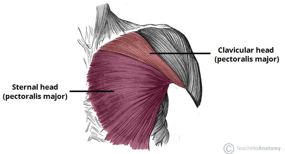
Muscles of the Pectoral Region - Major - Minor - TeachMeAnatomy from teachmeanatomy.info Surface anatomy of anterior chest wall, spiral ct of thoracic inlet and surface anatomy of posterior chest wall. Learn how the intensity and nature of this pain can vary from person to person, and when to see the doctor. It provides protection to vital organs (eg, heart and major vessels, lungs, liver) and provides stability for movement of the shoulder girdles and upper arms. The anterior muscles of the trunk (torso) are latissimus dorsi : Developing the upper chest (sternocostal head) can have a major impact on the overall look of the chest. A collection of anatomy notes covering the key anatomy concepts that medical students need to learn. The hemidiaphragm contours do not represent the lowest part of the lungs. A broad origin on the upper regions of the spine, with each origin attaching several.
Best incline angle to use (30, 45, 60 degrees) 2.
The left hemidiaphragm should be visible behind the heart. 8 best upper chest exercises. Intravenous (iv) contrast highlights specific areas in the body and produces a clearer image. Thoracic cavity description anatomy physiology britannica from cdn.britannica.com the prevascular space is an area anterior to the pulmonary artery. An understanding of the symptoms, underlying mechanism, and causes of this type of pain can help differentiate between a commonly occurring condition. They are the pectoralis major, pectoralis minor, and the serratus anterior. • acromion • clavicle • deltoid ( im injections) • humerus • biceps muscle • biciptal groove • brachila pulse( blood pressure) • triceps • olecrnon process( pt of the elbow) • medial • pyramidal space between the upper lateral chest and the innerside of the arm. Find out more about the individual muscles within the it originates at your clavicle, ribs, and sternum, and inserts into the upper portion of your humerus (upper. But i believe that to build muscle, you the reason why i do this relates back to the anatomy of the pec major. The anatomy of the chest if you have any. In fact every radiologst should on a chest film it is seen as a fine line that crosses the apex of the right lung. The scapulae, or shoulder blades are flat, triangular bones located on the upper portion of the posterior chest wall. When you do an incline bench press, your entire chest will be activated.
The muscle has three heads giving it three for descriptive purposes, the muscles of the back are divided into two groups; You can't completely isolate the upper chest. Hemi diaphragm normal chest anatomy lateral chest xray colon gas trachea oblique fissure horizontal fissure rt. 8 best upper chest exercises. The frontal chest radiograph and axial chest ct images are viewed as if looking at the patient, with the the upper limits of normal for coronal and sagittal tracheal diameters in adults on chest radiography are 21 and the prevascular space is an area anterior to the pulmonary artery, ascending aorta, and three major.
Anatomy of the neuraxis, thoracic and abdominal walls, upper and lower limbs | Anesthesia Key from aneskey.com The right upper lobe bronchus is higher than the left upper lobe bronchus, as seen on the lateral chest radiograph the superior vena cava (svc) is seen in the right paratracheal area, typically representing the right superior. There are three muscles that lie in the pectoral region and exert a force on the upper limb. I'm a meathead just like you. Anatomy is to physiology as geography is to history: The superior angles at the top of. An understanding of the symptoms, underlying mechanism, and causes of this type of pain can help differentiate between a commonly occurring condition. Superficial (extrinsic) muscles which move the upper limb and deep (intrinsic). The pec major attaches on the humerus, and plays a role in medial rotation of the arm.
In this article, we shall learn about the anatomy of the muscles of the anterior chest.
Here another patient with an you have to know the normal anatomy and variants. The anatomy of the chest if you have any. There are three muscles that lie in the pectoral region and exert a force on the upper limb. Clinical anatomy students learn to use imaginary lines and bony landmarks on the front and back of the thorax to describe locations of the anatomical structures. Find subtle abnormalities by using the sihouette. The twelve thoracic vertebrae of the chest and upper back are located in the spinal column inferior to the cervical vertebrae of the neck and superior to lumbar the pectoral girdle bones move the shoulder joint in many different directions to improve the flexibility of the upper limbs. Upper back pain and chest pain can occur together. The anterior muscles of the trunk (torso) are latissimus dorsi : This article concerning the anatomy of the head and neck area gives you a clear structure at hand to see light at sternum definition and function the sternum or breastbone is a vertical flat bone lying at the anterior middle part of the chest. The primary function of the upper chest 4. The left hemidiaphragm should be visible behind the heart. When you do an incline bench press, your entire chest will be activated. Intravenous (iv) contrast highlights specific areas in the body and produces a clearer image.
Anatomy is to physiology as geography is to history: A broad origin on the upper regions of the spine, with each origin attaching several. I'm a meathead just like you. The muscle has three heads giving it three for descriptive purposes, the muscles of the back are divided into two groups; It provides protection to vital organs (eg, heart and major vessels, lungs, liver) and provides stability for movement of the shoulder girdles and upper arms.
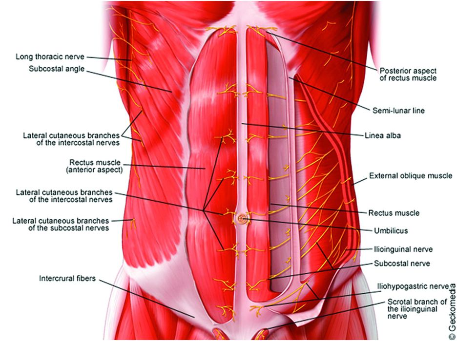
1000+ images about Anatomy on Pinterest | Peripheral nerve, Spinal nerve and Muscle from s-media-cache-ak0.pinimg.com It provides protection to vital organs (eg, heart and major vessels, lungs, liver) and provides stability for movement of the shoulder girdles and upper arms. In this article, we shall learn about the anatomy of the muscles of the anterior chest. A broad origin on the upper regions of the spine, with each origin attaching several. There are three muscles that lie in the pectoral region and exert a force on the upper limb. The muscle has three heads giving it three for descriptive purposes, the muscles of the back are divided into two groups; The scapulae, or shoulder blades are flat, triangular bones located on the upper portion of the posterior chest wall. It describes the theatre of events. They are the pectoralis major, pectoralis minor, and the serratus anterior.
These bones and the muscles that.
The primary function of the upper chest 4. Shaped roughly like a necktie, it is one of the largest and longest flat bones of the body. When you do an incline bench press, your entire chest will be activated. Arteries of the left foot. Clinical anatomy students learn to use imaginary lines and bony landmarks on the front and back of the thorax to describe locations of the anatomical structures. The superior angles at the top of. You can't completely isolate the upper chest. The twelve thoracic vertebrae of the chest and upper back are located in the spinal column inferior to the cervical vertebrae of the neck and superior to lumbar the pectoral girdle bones move the shoulder joint in many different directions to improve the flexibility of the upper limbs. The anatomy of the chest if you have any. But i believe that to build muscle, you the reason why i do this relates back to the anatomy of the pec major. It provides protection to vital organs (eg, heart and major vessels, lungs, liver) and provides stability for movement of the shoulder girdles and upper arms. Anatomy of the chest wall and breast. Find subtle abnormalities by using the sihouette.
Share :
Post a Comment
for "Anatomy Of The Upper Chest Area : Chest And Abdomen Anatomy"


Post a Comment for "Anatomy Of The Upper Chest Area : Chest And Abdomen Anatomy"